Development of Dendrimer-based Imaging Agents
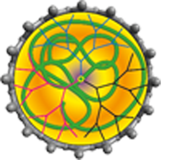
In collaboration with the group of Prof. J. Valliant and the Centre for Probe Development and Commercialization (CPDC), we have been developing macromolecular, dendrimer-based, radioimaging agents. Using 99mTc as the radionuclide and a bis(pyridyl) amine ligand at the dendrimer core, a series of high-generation aliphatic polyester dendrimers have been prepared and fully characterized. Their well-defined structure, high purity, and single-site radiolabeled structure produced clinically relevant imaging agents that could be evaluated in vivo using single-photon emission computed tomography (SPECT). This work represents early stages in the development of a dendrimer family that can house a therapeutic payload, be targeted to specific disease sites through peripheral modification, and be monitored in real-time upon injection into, perfusion through, and elimination from preclinical models and, ultimately, patients. The structures of high-generation dendrimers prepared thus far are shown below, and a time-lapse movie showing the biodistribution of the G-7 structure through the body of a live rat over the course of 15 minutes can be viewed in the video at the bottom of this page (an X-ray image is provided to give anatomical context to the images in the movie). 3-D tomographic images were recorded to prove that the dendrimer is eliminated through the kidneys and out the bladder. Current work is focused on the targeting of these macromolecules to specific disease sites.


Key Reference: Parrott, M. C.; Benhabbour, S. R.; Saab, C.; Lemon, J. A.; Parker, S.; Valliant, J. F.; Adronov, A. J. Am. Chem. Soc. 2009, 131, 2906-2916.